การค้นหาโดยอัตโนมัติของภาวะเบาหวานขึ้นจอตาจากภาพถ่ายจอประสาทตา
คณะเทคโนโลยีสารสนเทศและการสื่อสาร มหาวิทยาลัยมหิดล


การค้นหาโดยอัตโนมัติของภาวะเบาหวานขึ้นจอตาจากภาพถ่ายจอประสาทตา
คณะเทคโนโลยีสารสนเทศและการสื่อสาร มหาวิทยาลัยมหิดล
โครงการวิจัย:
การค้นหาภาวะเบาหวานขึ้นจอตาด้วยเทคนิคการประมวลผลภาพ
ผลงานวิจัย:
การค้นหาโดยอัตโนมัติของภาวะเบาหวานขึ้นจอตาจากภาพถ่ายจอประสาทตา
ผู้วิจัย:
ผศ. ดร.วรพันธ์ คู่สกุลนิรันดร์
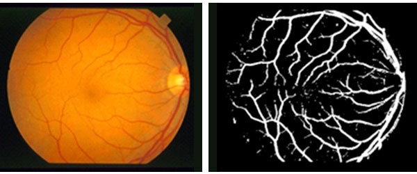
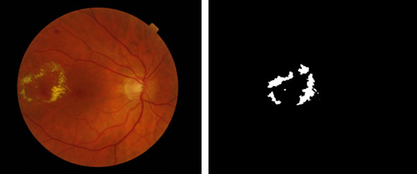
โครงการวิจัยนี้ มีวัตถุประสงค์หลักในการพัฒนาวิธีการและโปรแกรมคอมพิวเตอร์ ในการค้นหาและแบ่งระดับความรุนแรงของโรคเบาหวานขึ้นจอตาในภาพถ่ายจอประสาทตา ในรูปแบบอย่างเป็นอัตโนมัติ เพื่อที่จะสามารถช่วยในการคัดกรองภาพถ่ายจอประสาทตา สำหรับระดับความรุนแรงขั้นต้นที่สามารถรักษาให้หายขาดได้ หรือระดับความรุนแรงของโรคที่ต้องการการช่วยเหลืออย่างเร่งด่วน เพื่อไม่ให้เข้าสู่ภาวะการเสียการมองเห็น การมีโปรแกรมคอมพิวเตอร์ที่สามารถค้นหาโรค และวิเคราะห์ระดับความรุนแรงได้อย่างอัตโนมัติ จะสามารถช่วยให้การตรวจภาพถ่ายจอประสาทตาเข้าถึงคนไทยได้มากขึ้น โดยเฉพาะในบริเวณที่ห่างไกลสถานพยาบาล การพัฒนาในโครงการนี้ ใช้วิธีการประมวลภาพขั้นสูงในการแยกพื้นที่ภาพที่สามารถระบุระดับความรุนแรงได้ เช่น Microaneurysms, Hard Exudates และ Abnormal Blood Vessels ซึ่งสามารถใช้ต่อในการวิเคราะห์และประมวลผลการแยกระดับความรุนแรง โดยใช้การเรียนรู้ด้วยตัวเองของคอมพิวเตอร์ หรือ Machine Learning และในอีกส่วนของการพัฒนา คือ การใช้เทคนิคการเรียนรู้เชิงลึก เพื่อเรียนรู้ลักษณะของโรคเบาหวานขึ้นจอตาในแต่ละระดับ เพื่อให้สามารถแยกภาพถ่ายจอประสาทตาได้ วิธีการและโปรแกรมคอมพิวเตอร์ที่พัฒนาขึ้นมา ได้ทำการวัดประสิทธิภาพในทั้งมุมมองของการแยกพื้นที่ส่วนภาพในการระบุโรคออกจากพื้นที่ส่วนหลัง และการแยกระดับโรคของแต่ละภาพ ผลการทดลองได้ผลลัพธ์ที่น่าเชื่อถือด้วยความแม่นยำโดยเฉลี่ยมากกว่า 80% ในการแยกระดับของโรคในทุกระดับ


การเผยแพร่ผลงาน:
• W. Kusakunniran, Q. Wu, P. Ritthipravat, J. Zhang, Hard Exudates Segmentation based on Learned Initial Seeds and Iterative Graph Cut, Computer Methods and Programs in Biomedicine (CMPB), 158: 173-183, May 2018, DOI: 10.1016/j.cmpb.2018.02.011
• W. Kusakunniran, S. Kanchanapreechakorn, K. Thongkanchorn, Instance-based Learning for Blood Vessel Segmentation in Retinal Image, pages 111 – 118, Thailand, July 2019, International Conference on Computing and Information Technology (IC2IT), Advances in Intelligent Systems and Computing, Springer Nature Switzerland (IC2IT 2019, AISC 936), DOI: 10.1007/978-3-030-19861-9_11
• R. Kasantikul, W. Kusakunniran, Improving Supervised Microaneurysm Segmentation using Autoencoder-Regularized Neural Network, pages 553 – 559, Australia, December 2018, Digital Image Computing: Techniques and Applications (DICTA)
• W. Kusakunniran, Q. Wu, P. Ritthipravat, J. Zhang, Three-Stages Hard Exudates Segmentation in Retinal Images, pages 1 – 6, Thailand, October 2017, International Conference on Information Technology and Electrical Engineering (ICITEE)
รางวัลที่ได้รับ:
• ทุนพัฒนาศักยภาพในการทํางานวิจัยของอาจารย์รุ่นใหม่ สนับสนุนโดยสำนักงานกองทุนสนับสนุนการวิจัย
การติดต่อ:
ผศ. ดร.วรพันธ์ คู่สกุลนิรันดร์
คณะเทคโนโลยีสารสนเทศและการสื่อสาร มหาวิทยาลัยมหิดล